


Locations


Fax: (303) 762-9292
Fax: (303) 762-9292

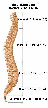
Understanding Spinal Anatomy: Regions of the Spine - Cervical, Thoracic, Lumbar, Sacral
The regions of the spine consist of the cervical, thoracic, lumbar, and sacral.
Cervical Spine
The neck region of the spine is known as the Cervical Spine. This region consists of seven vertebrae, which are abbreviated C1 through C7 (top to bottom). These vertebrae protect the brain stem and the spinal cord, support the skull, and allow for a wide range of head movement.
The first cervical vertebra (C1) is called the Atlas. The Atlas is ring-shaped and it supports the skull. C2 is called the Axis. It is circular in shape with a blunt tooth-like structure (called the Odontoid Process or dens) that projects upward into the Atlas. Together, the Atlas and Axis enable the head to rotate and turn. The other cervical vertebrae (C3 through C7) are shaped like boxes with small spinous processes (finger-like projections) that extend from the back of the vertebrae.
- Spinous Process
- Lamina
- Zygapophysial Joint (Facet)
- Posterior Tubercle
- Foramen
- Pedicle
- Body
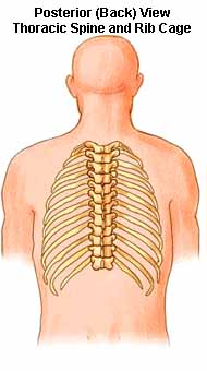
Thoracic Spine
Beneath the last cervical vertebra are the 12 vertebrae of the Thoracic Spine. These are abbreviated T1 through T12 (top to bottom). T1 is the smallest and T12 is the largest thoracic vertebra. The thoracic vertebrae are larger than the cervical bones and have longer spinous processes.
In addition to longer spinous processes, rib attachments add to the thoracic spine’s strength. These structures make the thoracic spine more stable than the cervical or lumbar regions. In addition, the rib cage and ligament system limit the thoracic spine’s range of motion and protects many vital organs.
- Spinous Process
- Lamina
- Zygapophysial Joint (Facet)
- Posterior Tubercle
- Foramen
- Pedicle
- Body
Lumbar Spine
The Lumbar Spine has 5 vertebrae abbreviated L1 through L5 (largest). The size and shape of each lumbar vertebra is designed to carry most of the body’s weight. Each structural element of a lumbar vertebra is bigger, wider and broader than similar components in the cervical and thoracic regions.
- Body
- Spinous Process
- Articular Process
- Transverse Process
- Foramen
- Pedicle
- Body
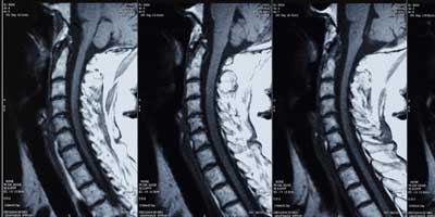
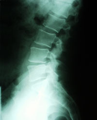 Side (lateral) x-ray view; lumbar spine
The lumbar spine has more range of motion than the thoracic spine, but less than the cervical spine. The lumbar facet joints allow for significant flexion and extension movement but limits rotation.
Sacral Spine
The Sacrum is located behind the pelvis. Five bones, abbreviated S1 through S5, fused into a triangular shape, form the sacrum. The sacrum fits between the two hip bones connecting the spine to the pelvis. The last lumbar vertebra (L5) articulates (moves) with the sacrum.
Immediately below the sacrum are five additional bones, fused together to form the Coccyx (tailbone).